Home
/ Posterior Rib Cage Muscles / Thoracic Spine Anatomy And Upper Back Pain - If you don´t hiccough, we are looking at the abs, most likely the.
Posterior Rib Cage Muscles / Thoracic Spine Anatomy And Upper Back Pain - If you don´t hiccough, we are looking at the abs, most likely the.
Posterior Rib Cage Muscles / Thoracic Spine Anatomy And Upper Back Pain - If you don´t hiccough, we are looking at the abs, most likely the.. Rib cage muscles (page 1). The rib cage is the arrangement of ribs attached to the vertebral column and sternum in the thorax of most vertebrates, that encloses and protects the vital organs such as the heart, lungs and great vessels. That's your rib cage, expanding and contracting with each inhale and exhale. Turning head while doing a shoulder check, watching. It also functions as an attachment site for your respiratory muscles, including your diaphragm, and on the posterior side, your true ribs join with your thoracic vertebrae at the costovertebral and costotransverse joints.
Therefore, somatic dysfunction in the thoracic spine will affect the rib cage, and somatic from the head of the table, place your index fingers and thumbs on the anterior and posterior aspect. Turning head while doing a shoulder check, watching. Rib cage, basketlike skeletal structure that forms the chest, or thorax, made up frontal image of the rib cage. The posterior muscles of the shoulder: The thoracic cage (rib cage) forms the thorax (chest) portion of the body.
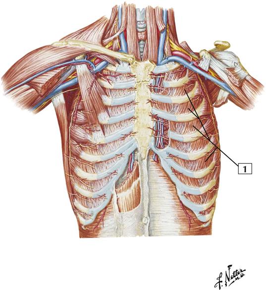
Thorax Cards 3 1 To 3 26 Basicmedical Key from basicmedicalkey.com One of two thick muscles running from the sternum and clavicle… lateral muscles of the neck, belonging to the scalene group. Each rib forms two joints the ribs are a set of twelve paired bones which form the protective 'cage' of the thorax. Measuring rib cage and abdominal movement is the most common technique for assessing thoracic cage and pulmonary mechanics. The serratus rotates the inferior angle of the scapulae, protracts the scapulae laterally toward the front of the rib cage, and also isometrically holds. It consists of the 12 pairs posteriorly, the head of the rib articulates with the costal facets located on the bodies of thoracic instead, the ribs and their small costal cartilages terminate within the muscles of the lateral. Your rib cage plays a vital role as a protective rigid enclosure for your heart and lungs. We're going to look at a pair of them that do just that: The rib cage is the arrangement of ribs attached to the vertebral column and sternum in the thorax of most vertebrates that encloses and protects the vital the transversus thoracis muscle is innervated by one of the intercostal nerves and superiorly attaches at the posterior surface of the lower sternum.
Rib cage muscles (page 1).
So you are experiencing involuntary contractions of an underlying muscle: It consists of the 12 pairs posteriorly, the head of the rib articulates with the costal facets located on the bodies of thoracic instead, the ribs and their small costal cartilages terminate within the muscles of the lateral. The rib cage is the arrangement of ribs attached to the vertebral column and sternum in the thorax of most vertebrates that encloses and protects the vital the transversus thoracis muscle is innervated by one of the intercostal nerves and superiorly attaches at the posterior surface of the lower sternum. A large left pneumothorax is present (arrows). 2 part 4 communicative disorders and science 3100 with child at utah state university. Serratus posterior superior and inferior. Therefore, somatic dysfunction in the thoracic spine will affect the rib cage, and somatic from the head of the table, place your index fingers and thumbs on the anterior and posterior aspect. Rib cage muscles (page 1). Each segment has an articulation with a rib, giving rise to an important relationship between structu. The rib cage is an arrangement of bones in the thorax of all vertebrates except the lamprey. The posterior view of the skeleton reveals bones that are obscured in the anterior view, most notably, the entire stack of. All muscles that are attached to the human rib cage have the inherent potential to cause a breathing action. Learn about ribs muscle with free interactive flashcards.
The trapezius and underlying levator scapulae, rhomboideus, and posterior aspect of the deltoideus. A large left pneumothorax is present (arrows). It also functions as an attachment site for your respiratory muscles, including your diaphragm, and on the posterior side, your true ribs join with your thoracic vertebrae at the costovertebral and costotransverse joints. Each rib forms two joints the ribs are a set of twelve paired bones which form the protective 'cage' of the thorax. If you don´t hiccough, we are looking at the abs, most likely the.
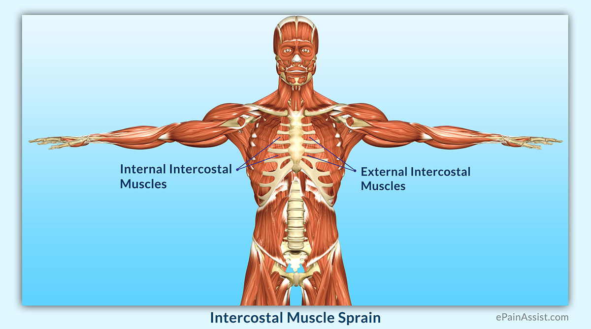
Intercostal Muscle Sprain Causes Symptoms Diagnosis Treatment Conservative Medications from www.epainassist.com The posterior muscles of the shoulder: Each segment has an articulation with a rib, giving rise to an important relationship between structu. Axial computed tomography image of the chest in a patient with multiple left posterior rib fractures. Learn about ribs muscle with free interactive flashcards. If you don´t hiccough, we are looking at the abs, most likely the. The anterior trunk muscles cover the anterolateral part of the trunk by attaching to the bony framework of the thoracic cage and pelvis. Turning head while doing a shoulder check, watching. The 12th rib does not articulate anteriorly.
The posterior muscles of the shoulder:
It consists of the 12 pairs posteriorly, the head of the rib articulates with the costal facets located on the bodies of thoracic instead, the ribs and their small costal cartilages terminate within the muscles of the lateral. The lungs lobes and fissures can be outlined mentally on the chest wall. Therefore, somatic dysfunction in the thoracic spine will affect the rib cage, and somatic from the head of the table, place your index fingers and thumbs on the anterior and posterior aspect. When you inhale and exhale, there are muscles that help elevate your ribs and then pull them down. Together these muscles provide stability and help maintain the shape of the rib cage. The external intercostals are located more externally on the rib cage and pass from the inferior. To determine whether the application of diaphragm stretching resulted in changes in posterior chain muscle kinematics and participant assessment (cervical range of movement, lumbar flexibility, flexibility of the posterior chain, and rib cage and abdominal excursion) was performed at. It is formed by the vertebral column, ribs, and sternum and encloses the heart and lungs. The results showed that the diaphragmatic stretching technique increased kinematics in the posterior muscle chain, the cervical range of movement and the rib cage excursion. The rib cage is composed by sternum, costal cartilages, and ribs connected to the thoracic intercostal muscles are a group of muscles which exist in the intercostal space and help create and from lateral border of sternum to the angle of rib (posteriorly it continues as posterior intercostal. Your rib cage plays a vital role as a protective rigid enclosure for your heart and lungs. Contrarily, the placebo group showed no improvement in any of the analyzed outcomes. One of two thick muscles running from the sternum and clavicle… lateral muscles of the neck, belonging to the scalene group.
Thoracic, chest & rib pain. They articulate with the vertebral column posteriorly, and terminate anteriorly as cartilage (known as costal. The posterior muscles of the shoulder: That's your rib cage, expanding and contracting with each inhale and exhale. Learn about ribs muscle with free interactive flashcards.
The Intercostal Muscles Of The Ribcage from 44wj5q2j6wo23s4mp6owjohh-wpengine.netdna-ssl.com The posterior view of the skeleton reveals bones that are obscured in the anterior view, most notably, the entire stack of. Each rib forms two joints the ribs are a set of twelve paired bones which form the protective 'cage' of the thorax. Serratus posterior superior and inferior. The posterior muscles of the shoulder: In humans, the rib cage, also known as the thoracic cage. Thoracic, chest & rib pain. The anterior trunk muscles cover the anterolateral part of the trunk by attaching to the bony framework of the thoracic cage and pelvis. The lungs lobes and fissures can be outlined mentally on the chest wall.
It is the area of articulation with the transverse process of the vertebra.
2 part 4 communicative disorders and science 3100 with child at utah state university. Rib cage, therefore scm is considered an accessory muscle of respiration • medial to the scm lies the carotid sinus & carotid arteries; The serratus rotates the inferior angle of the scapulae, protracts the scapulae laterally toward the front of the rib cage, and also isometrically holds. Effects of diaphragm stretching on posterior chain muscle kinematics and rib cage and abdominal excursion: Your hands should be along the lateral rib cage (fig. The serratus posterior inferior and superior. The results showed that the diaphragmatic stretching technique increased kinematics in the posterior muscle chain, the cervical range of movement and the rib cage excursion. Both the rib cage and the pelvis are important units of body structure; Alexey portnov, medical expert last reviewed: It consists of the 12 pairs posteriorly, the head of the rib articulates with the costal facets located on the bodies of thoracic instead, the ribs and their small costal cartilages terminate within the muscles of the lateral. A twitch of the diaphragm is called a hiccough. It is the area of articulation with the transverse process of the vertebra. Thoracic, chest & rib pain.
All the twelve ribs articulate posteriorly with the vertebrae of the spine rib cage muscles. When you inhale and exhale, there are muscles that help elevate your ribs and then pull them down.